Augusta University
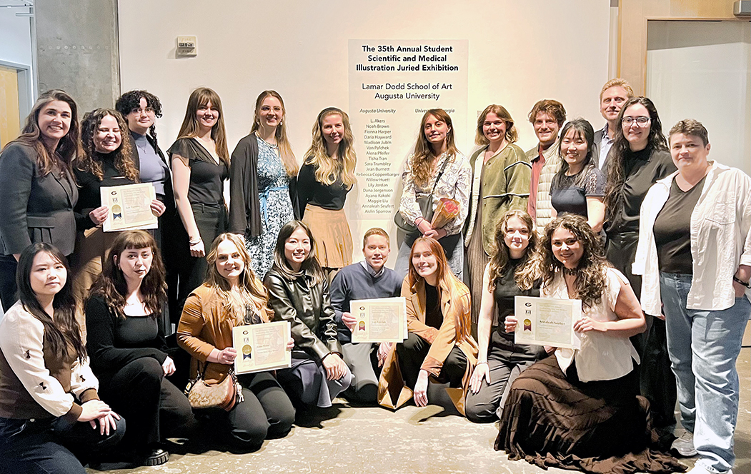
Medical illustrators at Augusta University and the University of Georgia celebrated their long-standing partnership with the 35th annual Scientific and Medical Illustration Exhibit in Athens, Georgia as the schools aim to highlight and celebrate the art forms that move natural and medical science forward.
Each year, the exhibition showcases the scientific and medical storytelling of Georgia’s Scientific Illustration Undergraduate Program and AU’s Medical Illustration graduate program, which is located in the College of Allied Health Sciences. The exhibits are displayed at the Lamar Dodd School of Art.
This year’s exhibit showcased the vital role of visual communication in transforming complex scientific and medical information into accessible and engaging artwork. The show demonstrated how illustration serves as both an educational tool and a bridge between scientific knowledge and the public by featuring anatomical studies, patient education visuals and natural science illustrations.
Eighteen students from AU and 16 from UGA participated. This year’s exhibition was organized by assistant professor Amanda Manowski, who is the Scientific Illustration section chair at UGA and is an alumna of the AU graduate program.
Five professional medical illustrators evaluated the submissions, focusing on the clarity and effectiveness of each work’s scientific narrative.
“The judges remarked on the high quality of the work presented and noted how challenging the final award decision was,” said Amanda Behr, PhD, department chair and professor. “I am incredibly proud of all our students, who served as exemplary ambassadors of our program and truly deserve recognition for their hard work.”
Four students honored
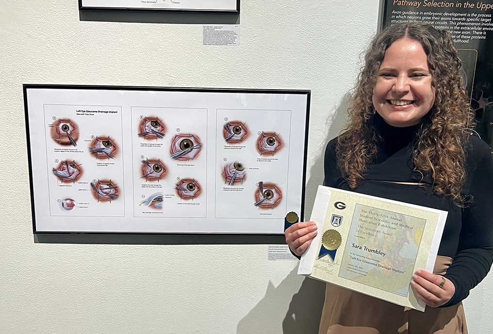
Stenstrom Award of Excellence: Sara Trumbley, Left Eye Glaucoma Drainage Implant. Trumbley’s illustration is an accurate and clear depiction of an ophthalmic surgical implant with a strong narrative flow. Her work reflects the program’s rigorous training in surgical illustration and benefits from the close collaboration with Wellstar MCG Health, where students observe surgical procedures and work with surgeons in an immersive educational experience.
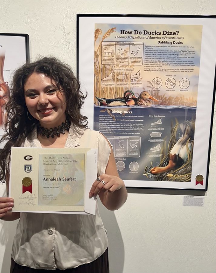
Award of Merit: Annaleah Seufert, How Do Ducks Dine. Seufert’s illustration offers an engaging, well-researched look at avian feeding anatomy and behavior, blending scientific accuracy with strong visual storytelling. This piece was a community outreach project in partnership with the Georgia Department of Natural Resources.
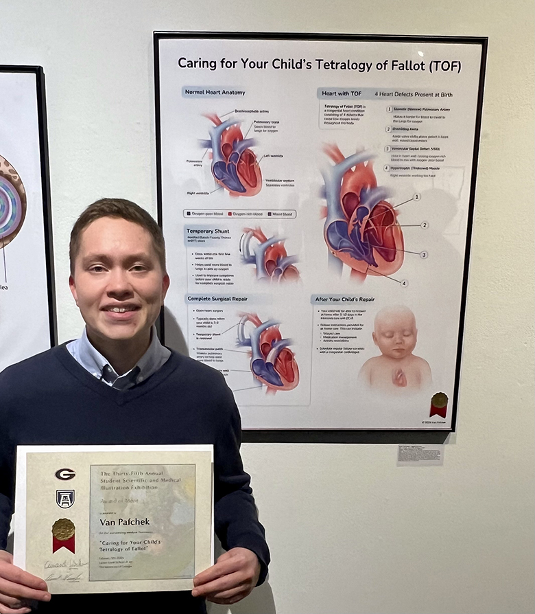
Award of Merit: Van Pafchek, Caring for Your Child’s Tetralogy of Fallot. Pafchek’s work offers a compassionate and effective educational resource, translating the complexity of a congenital heart condition into visuals that support both patient understanding and clinical communication. This piece highlights the essential role medical illustrators play in advancing health literacy.
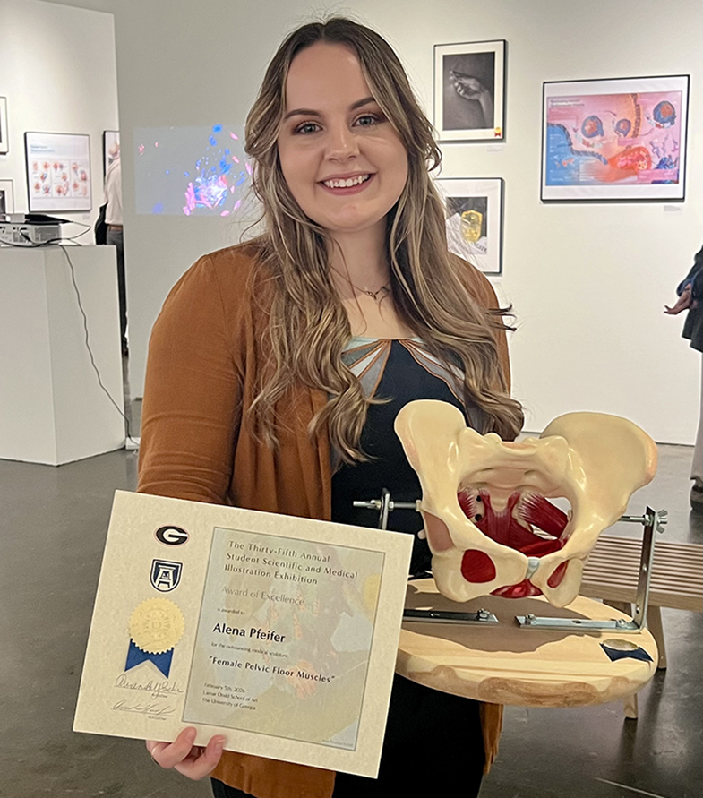
Award of Excellence: Alena Pfeifer, Female Pelvic Floor Muscles. Pfeifer’s 3D anatomical reconstruction of the female pelvic floor highlights the capability of three-dimensional modeling to communicate complex anatomical structures with exceptional clarity. Created in the Medical Sculpture Core Lab using digital software and 3D printing, the piece demonstrates the anatomical form and function of the pelvic floor through meticulous modeling of accurate anatomical detail.
The 36th annual Student Scientific and Medical Illustration Juried Exhibition will be supported by AU’s College of Allied Health Sciences and the David Mascaro Fund. For support opportunities, email Amanda Behr.