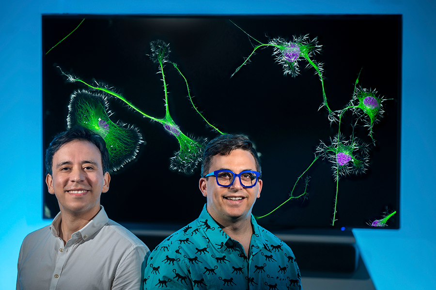
It’s a picture that is equal parts art and science.
At first glance, the green and magenta hues might trick the eye into seeing Venus fly traps or plankton, but the image actually shows differentiated mouse brain tumor cells, highlighting the actin cytoskeleton, microtubules and nuclei of the cells.
Even though the image won a photography contest, it wasn’t made exclusively for art as it’s part of a research study Bruno Cisterna, PhD, submitted to the Journal of Cell Biology.
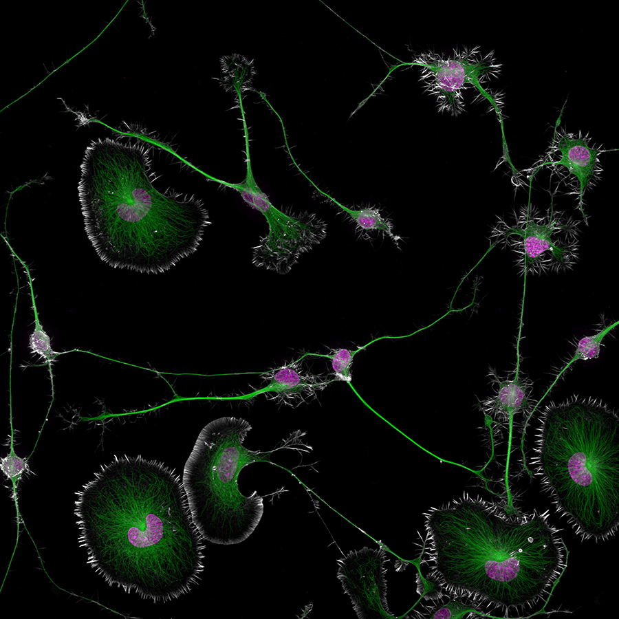
Cisterna, a research scientist in the Department of Neuroscience and Regenerative Medicine in Augusta University’s Medical College of Georgia, created the image with the help of Eric Vitriol, PhD, director of the Neuroscience Graduate Program, and the duo submitted the image and took home top honors in the 50th anniversary of Nikon Small World Photomicrography Competition. The competition showcases the beauty and complexity of life as seen through the light of the microscope. In total, Nikon Small World recognized 87 photos out of thousands of entries from scientists and artists across the globe.
“This has been something I’ve had my eye on doing for about 15 years, but I never had a picture I thought was good enough,” Cisterna said. “This year we finally found something that was both an interesting image and an interesting research subject. It’s an incredible contest that highlights the beauty of photomicrography but also inspires continued exploration and innovation in the field.”
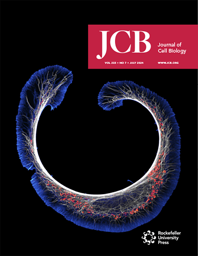
their image “Early stage of mouse
neuroblastoma cell differentiation (actin,
microtubules, and mitochondria).”
Cisterna and Vitriol also took home 20th with their image showing early-stage mouse neuroblastoma cell differentiation (actin, microtubules and mitochondria). That image was the cover photo of the July 2024 cover of the Journal of Cell Biology, and it was the one Cisterna thought would be the winner.
The final image took four months of work to get everything just right and is a composite of over 860 individual images. But true to his artistic eye, Cisterna feels he has other images that are better.
While there is certainly an art to creating images of this caliber, it’s also science. Cisterna noted the image needs to be clear so scientists can better study the parts of the cells. If you have a fuzzy or out-of-focus image, you cannot study what is happening within the cells.
“Bruno just had a paper published in the Journal of Cell Biology, and he was working on putting cover images together,” Vitriol said. “When we take images on the microscope, they’re usually created to gather data, not to make something that looks beautiful. But for the cover, there’s a little bit more of an artistic aspect to it.
“Bruno has the soul of a poet combined with being a really fantastic scientist, and that’s the perfect combination to create an image that looks amazing and tells a story about the study it represents,” Vitriol added.
Over about three months, Cisterna focused on getting the staining just right so he could have clear visibility of the cells. This included waiting five days after each stain to allow the cells to differentiate, then working to find the right field of view to show the differentiated and non-differentiated cells interacting, a process that took three hours of patience and precise observation under the microscope.
Over the span of a week, Cisterna worked on creating samples that would allow him to capture the image he had in mind. During that time, he focused on capturing multi-color, super-resolution images of the cytoskeleton.
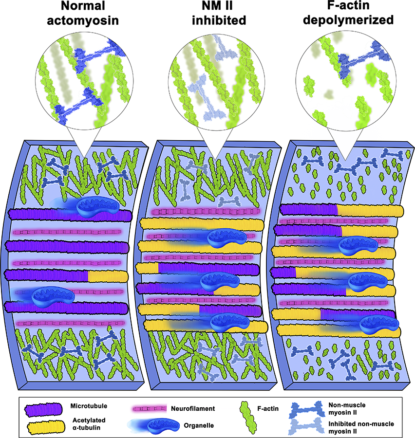
responses in other cytoskeleton components,
which in turn significantly alter cellular function.
Cisterna’s research, which he has been working on for three years, is based around understanding the root cause of neurodegenerative diseases like Alzheimer’s or ALS.
Profilin 1, a vital protein for cellular architecture, was found to play a primary role during Cisterna’s study. This molecule safeguards microtubule pathways, ensuring smooth cellular transport. Disruptions to PFN1 or its associated mechanisms can derail these highways, triggering cell damage reminiscent of neurodegenerative disorders.
“One of the main problems with neurodegenerative diseases is that we don’t fully understand what causes them,” said Cisterna. “To develop effective treatments, we need to figure out the basics first. Our research is crucial for uncovering this knowledge and ultimately finding a cure. Differentiated cells could be used to study how mutations or toxic proteins that cause Alzheimer’s or ALS alter neuronal morphology, as well as to screen potential drugs or gene therapies aimed at protecting neurons or restoring their function.”
Discoveries at Augusta University are changing and improving the lives of people in Georgia and beyond. Your partnership and support are invaluable as we work to expand our impact.
 Augusta University
