Augusta University
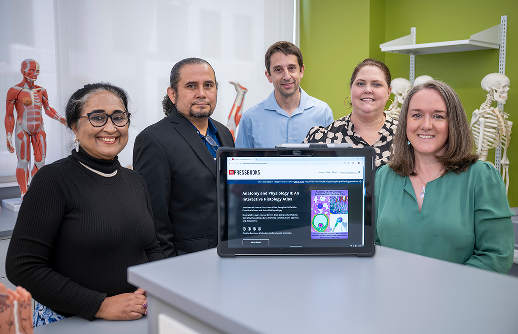
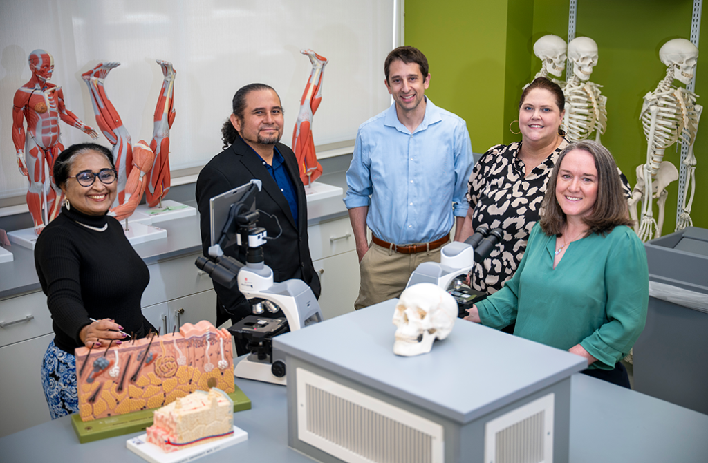
A group of five faculty members from Augusta University’s College of Science and Mathematics and the Medical College of Georgia has developed a way around the expense of college textbooks by creating interactive, digital textbooks that cover the materials while allowing students to keep a little more money in their pockets.
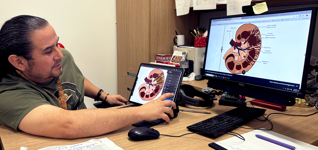
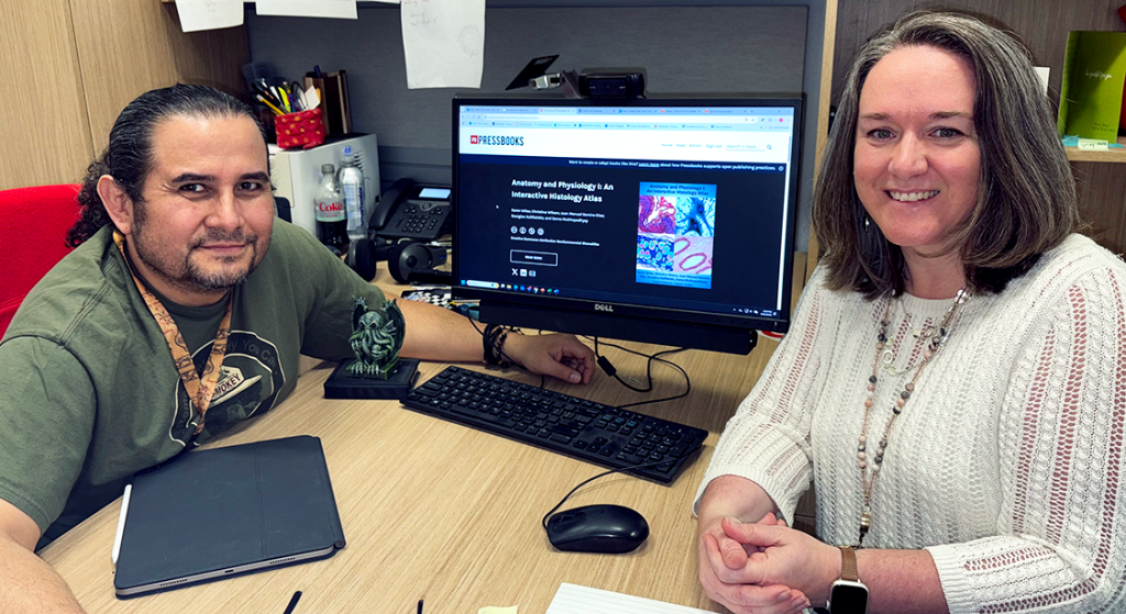
“The story started one day when I forgot to get my PowerPoints for my lab, so I had to draw the pictures on the board, and then I used that as my cues to teach my lab,” said Juan Manuel Ramiro Diaz, PhD, a lecturer in the Department of Biological Sciences at AU. “It was Anatomy and Physiology I, and, since then, I started using them. And then Dr. Wiles was the one who came up with the idea.”
“I hated erasing his drawings for the week,” said Karen Wiles, PhD, an assistant professor in the Department of Physiology at MCG. “He would go in on Friday and draw the pictures for the whole next week – he and Dr. Kallifatidis would fill the whiteboards for both Anatomy and Physiology I and Anatomy and Physiology II, and every Friday we’d have to erase all their beautiful art.”
But what if the art didn’t have to be erased?
Ramiro Diaz and Wiles discussed the difference the drawings had made in his lab – the realistic depictions compared to the traditional textbook photos and the students’ positive responses to them.
“A lot of our pre-health majors have to take A&P I and II, and everything looks like speckles of pink and purple and blue, and the students have a hard time figuring out, ‘What am I supposed to be looking at? Am I seeing what the professor wants me to see?’” Wiles said. “So these drawings would give them a little bit of the professor’s imagination to help connect what they should be seeing with what they’re actually seeing.”
Ramiro Diaz credits Wiles for the idea to translate not just the drawings but also the entire curriculum into a digital textbook format for the students to access whenever they need it.
This will hopefully solve a few issues students commonly face with the courses. For starters, faculty members who have been studying the subjects for years are used to looking into a microscope and being able to easily identify the structures they’re seeing.
“But for a student’s first time looking in the microscope, everything looks the same, so it was more like giving them an idea of what we see,” Ramiro Diaz said.
Another issue the drawings brought clarity to was how the pictures in traditional textbooks don’t always match up with what a student actually sees when they look into a microscope.
“The pictures that the old textbooks chose were super high magnification and were not representative of the slides that the students had at all,” Wiles explained. “So if the students had a microscope that could maybe get up to 400 times magnification, the picture in the textbook might be far higher than that. Just super unhelpful for the students.”
In 2022, the plan to design a new and improved, highly interactive, free online textbook was set into motion.
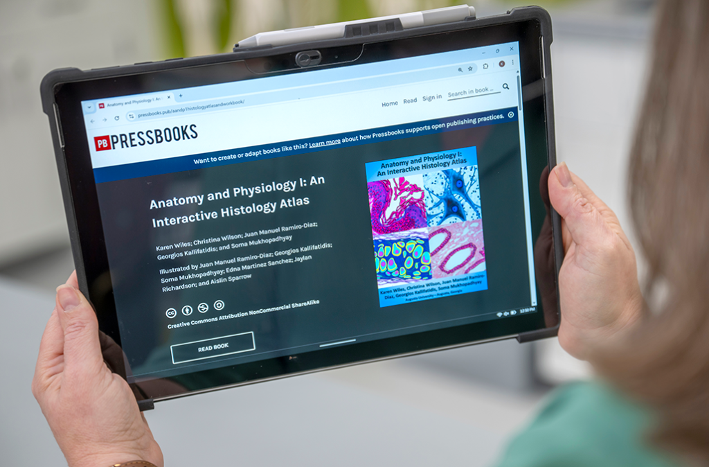
The books, titled Anatomy and Physiology I: An Interactive Histology Atlas and Anatomy and Physiology II: An Interactive Histology Atlas, were created and written by Ramiro Diaz and Wiles, along with their colleagues Georgios Kallifatidis, PhD; Soma Mukhopadhyay, PhD; and Christina Wilson, PhD, using grant funding awarded by the University System of Georgia’s Affordable Learning Georgia initiative.
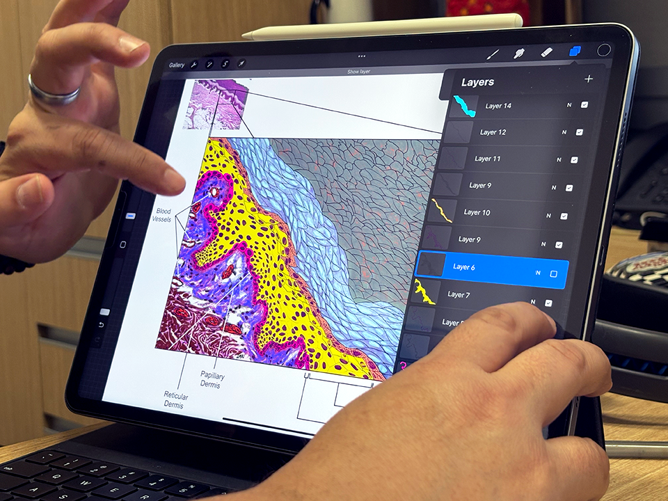
They applied for a Textbook Transformation Grant for both books and were awarded enough funding to purchase iPads so the faculty members in charge of illustrating could render the microscope photos in the books.
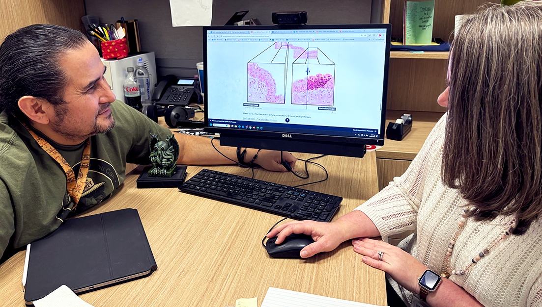
“When we created this book, we took pictures of the student slides. We didn’t pick all the textbook perfect images; we didn’t pick the most perfect representation,” Wiles said. “We picked what the students actually see and their slides that are commercially available, that every lab in the USG is using, because we all purchase from pretty much the same companies. So hopefully this textbook is usable and approachable to students no matter where they’re taking the class.”
The grant cycle for the second book is about to end, and, this time around, they were able to use some of the money to bring students on board.
“A lot of people don’t know this, but Augusta University is one of only four graduate-level medical illustration programs in the entire country, and a lot of our A&P students are aspiring medical illustrators,” Wiles said. “So, we hired a couple of them to create drawings that are going to go into our A&P I and II books.”
Five AU undergraduate students currently have illustrations published in the textbooks.
“Contributing to the atlas’ illustrations allowed me to combine my passions for anatomy and art. Being a part of this project has been both fun and rewarding, knowing that my work can support others in their learning,” said Karis Le, a third-year kinesiology student.
While countless students have benefited from the work of Le and her counterparts, the illustrators also have gained invaluable knowledge and experience that could propel their collegiate and professional careers forward.
“Working on this project was such an exciting experience! Having the opportunity to illustrate and publish my piece in the histology atlas was both an honor and a privilege. I gained so much experience and insight by collaborating with all the professors involved,” said Zoey Collins, a second-year cell and molecular biology student.
Besides the fact that the new books help the students learn in a more efficient way, one of the biggest takeaways from the project is the amount of money it’s estimated to have saved them. The students are no longer required to purchase the $100 lab manual for the courses, only the lecture textbook.
A&P I textbook savings began in the spring of 2024, and an estimated $38,596 has been saved by the students so far. A&P II textbook savings began in the spring of 2025, and an estimated $9,495 has been saved so far.
The textbooks are open access and are published in OER Commons and the University System of Georgia library, allowing students on both the Summerville and Health Sciences campuses access to the books. In just over a year, they’ve had more than 56,000 total viewers.
“For the grant, we were not required to have any sort of proof of concept. We had a proposal and framework of the type of product we wanted to create. And it was also a really important part of the initiative to decrease the cost for students and remove that barrier to access for educational materials,” Wiles said. “We were awarded that in November of 2023, and then the textbook went live in January 2024.”
It took a few months to start writing and adding the images to the books.
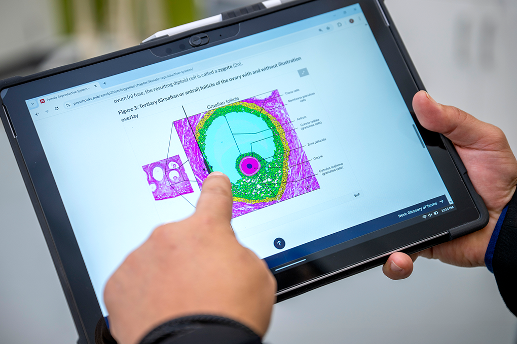
“In many cases in the book, there’s lots of different examples of what a tissue type could look like, and we wanted students to have a wide, broad range of reference images, so that was the bulk of our early work,” Wiles said.
The books are still being refined, and faculty members with access can make edits as often as they’d like. Since they are interactive, students can use slider tools on the images to quiz themselves and see before-and-after renderings of the images.
The platform works on all devices, so it looks the same on an iPhone as it does on a Windows PC.
Based on a survey the faculty members conducted, the books have been a hit.
“The response was overwhelmingly positive from the students, and we’ve implemented some of the changes and feedback they’ve given us,” Wiles said. “They asked for some practice quizzes, so we built in some practice quizzes into the A&P I book, where the students can practice what a lab practical would look like. They have to identify a tissue type and type in their answer. We were able to add those types of elements to the book in response to that feedback.”
The team has started to move on from drafting the histology of tissues, working on illustrating the organs themselves.
“Once you start with this thing, then you start coming up with more and more ideas, and then you can modify it by yourself,” Ramiro Diaz said. “The sky’s the limit.”