 Augusta University
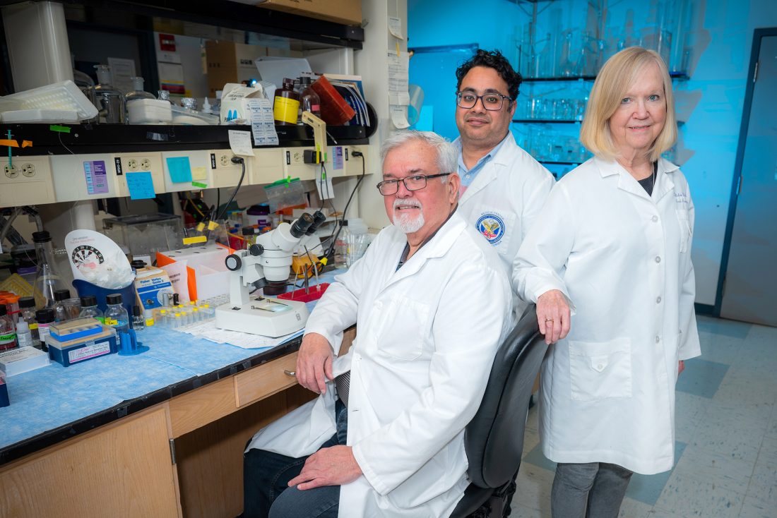
 Tim Rausch
Researchers who previously linked a cholesterol-processing pathway to retinal damage in premature infants have extended their work to diabetic retinopathy, finding that the same pathway may contribute to inflammation, blood vessel damage and nerve cell loss in the eye.
The latest study from scientists at the Medical College of Georgia at Augusta University examined what happens when cholesterol esters, a storage form of cholesterol, accumulate in the retina of a diabetic eye. The study was supported by a National Eye Institute R01 grant, awarded to Modesto A. Rojas, MD, and Ruth B. Caldwell, PhD.
The research, published in July in Investigative Ophthalmology & Visual Science, found that blocking the enzyme responsible for producing those cholesterol esters reduced several signs of retinal damage in diabetic mice.
“Normally, cells store excess cholesterol safely inside lipid droplets as esterified cholesterols,” said lead author Syed Adeel H. Zaidi, PhD, a research scientist in the Vascular Biology Center and Culver Vision Discovery Institute at MCG. “But we have found that too much esterified cholesterol is toxic and it causes immune cells, macrophages and microglia, to become activated and start causing inflammation, which is bad for the retina.”
The study builds on research the team published in 2023 on retinopathy of prematurity, an eye disease that can affect premature infants. That work found that the same cholesterol pathway contributed to inflammation and the growth of abnormal blood vessels. The new publication applies that discovery to diabetic retinopathy, a chronic disease that damages an already developed retina.
When cholesterol storage goes wrong
The enzyme at the center of the research is known as ACAT1. Researchers also refer to it as SOAT1. The enzyme helps cells convert free cholesterol and fatty acids into cholesterol esters.
That process usually helps cells manage cholesterol and maintain normal functions. In diabetes, however, the enzyme appears to become more active as the retina accumulates excessive cholesterol esters.
Macrophages are immune cells that remove damaged material and help regulate inflammation. Microglia perform similar work in the retina and other parts of the central nervous system. When these cells remain activated, they can continue releasing inflammatory signals that injure nearby tissue. The researchers proposed that excessive cholesterol ester formation helps sustain that response in diabetic retinopathy.
In their animal model, the scientists tested K604, an experimental compound that blocks ACAT1/SOAT1 and is being explored in cancer and cardiovascular disease research. The treatment reduced features of diabetic retinopathy without lowering blood glucose levels.
“Most of the research on diabetic retinopathy is about managing glucose levels” said Rojas, a vascular biologist in the MCG Department of Pharmacology and Toxicology and Vascular Biology Center. “What we found is that when we gave this drug, it did not decrease blood glucose levels. It was working independently by decreasing the levels of cholesterol esters.”
That distinction could give researchers a way to address retinal damage alongside the medications patients already use to manage diabetes.
Protecting blood vessels and nerve cells
Diabetic retinopathy has often been viewed primarily as a blood vessel disease. The condition can weaken retinal vessels, allowing material from the bloodstream to leak into surrounding tissue.
The disease also affects the neural retina, including cells that help capture, process and transmit visual information.
“Hypercholesterolemia has long been associated with inflammation and vascular dysfunction. However, the mechanisms linking altered cholesterol metabolism to diabetic retinal injury remain incompletely understood,” said Caldwell, a professor of cellular biology and anatomy, vascular biology and ophthalmology at MCG and a co-corresponding author of the paper.
In the preclinical study, blocking the cholesterol-processing enzyme reduced oxidative stress, a process in which unstable molecules damage cells. The treatment also reduced inflammatory signals and limited the abnormal attachment of white blood cells to retinal blood vessels.
The compound reduced vascular leakage and prevented an increase in acellular capillaries, damaged vessels that have lost the cells required to function. It also limited the loss of retinal ganglion cells, which help transmit visual information from the eye to the brain.
Tests of retinal function showed that the treatment preserved electrical responses during early and later stages of diabetes. Measured visual acuity improved significantly during the later stages, while the early-stage results showed a positive trend, it did not reach statistical significance.
Together, the findings suggest that the cholesterol pathway contributes to vascular and neural damage rather than affecting only one portion of the retina.
The findings support the biological relevance of the pathway, but they do not establish K604 as an effective treatment for patients. The compound has been evaluated only in preclinical studies.
“We are the first to report the role of the enzyme and esterified cholesterol in the pathogenesis of diabetic retinopathy,” Zaidi said.
Building the next stage of research
The next phase will focus on how the cholesterol pathway affects immune cells in the retina and how blocking it produces the protective effects reported in the study.
“The next step is to understand, from a mechanistic standpoint, how the drug is working,” Zaidi said.
Considerable development work remains before the findings could lead to a treatment for patients. Researchers would need to confirm the results in additional disease models and determine an appropriate treatment method, dose and long-term safety profile.
“Our goal is to provide strong preclinical data so that we can evaluate both the target and the drug itself,” Rojas said. “Our ultimate goal is to determine whether this drug can be repurposed for diabetic retinopathy in the future.”


 Jennifer Hilliard Scott
![Participants listen and engage during the Connected for Life Open-House event. [Aaron Smith/Augusta University]](https://jagwire.augusta.edu/wp-content/uploads/sites/15/2026/10/DSC_9186-332x249.jpg)
 Aaron Smith
 Paige Tucker
 Valerie Emerick